Click on the links below to find out more
DDH-Developmental Dislocation (Dysplasia)/Congenital Hip Dislocation
DESCRIPTION
In all cases of DDH, the socket (acetabulum) is shallow, meaning that the ball of the thighbone (femur) cannot firmly fit into the socket. Sometimes, the ligaments that help to hold the joint in place are stretched. The degree of hip looseness, or instability, varies among children with DDH.
CONDITION
Dislocated
In the most severe cases of DDH, the head of the femur is completely out of the socket.
Dislocatable
In these cases, the head of the femur lies within the acetabulum, but can easily be pushed out of the socket during a physical examination.
Subluxatable
In mild cases of DDH, the head of the femur is simply loose in the socket. During a physical examination, the bone can be moved within the socket, but it will not dislocate.
Causes
DDH tends to run in families. It is also predominant in:
- Girls
- First-born children
- Babies born in the breech position (especially with feet up by the shoulders).
The American Academy of Pediatrics now recommends ultrasound DDH screening of all female breech babies. - Family history of DDH (parents or siblings)
- Oligohydraminos (low levels of amniotic fluid)
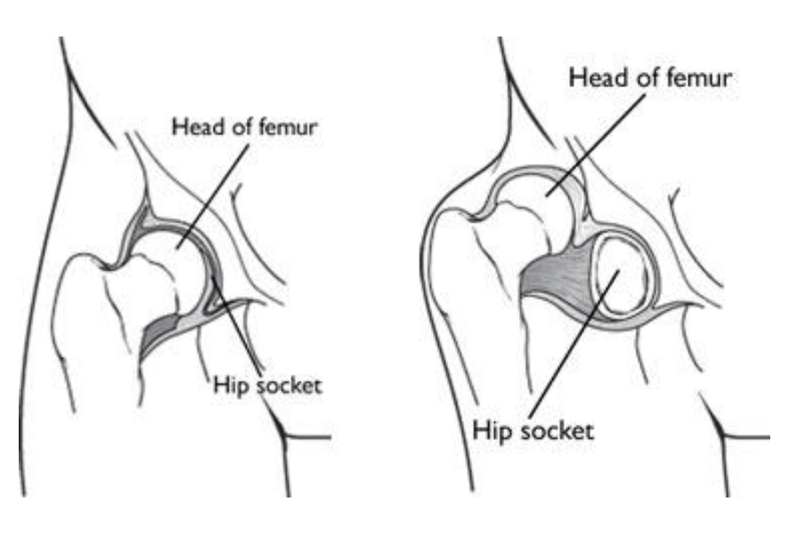
(Left) In a normal hip, the head of the femur fits firmly inside the hip socket. (Right) In severe cases of DDH, the femur is completely out of the hip socket (dislocated).
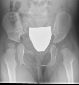
X Ray Image showing a dysplastic left hip – note the shallow socket, and angled roof(line)
Symptoms
Some babies born with a dislocated hip will show no outward signs.
Contact your GP or Paediatrician if your baby has:
- Legs of different lengths
- Uneven skin folds on the thigh
- Less mobility or flexibility on one side
- Limping, toe walking, or a waddling, duck-like gait

Dr David Slattery
FRACS MBBS (Hons) LLB FAOrthA
Dr David Slattery is an orthopaedic surgeon based in Melbourne with over 10 years of experience, with a special focus on hip and knee joint preservation and replacement. With qualifications in both medicine and law, he brings a unique and comprehensive approach to patient care. His surgical techniques are minimally invasive and evidence-based, designed to reduce pain and enhance recovery.
Trained in leading institutions across Europe and the USA, Dr Slattery offers advanced treatments for a wide range of joint conditions. He is deeply committed to patient outcomes and takes pride in tailoring treatment plans to each individual. Whether you’re an athlete or seeking relief from chronic joint pain, his goal is to restore function and improve your quality of life.


