Click on the links below to find out more


Patella Stabilisation
OVERVIEW
Patella stabilisation is a broad term to describe surgery that is used to stabilise (prevent dislocation) of the kneecap (patella). There are many options for this type of surgery, which can include both bone and soft tissue procedures or a combination of both.
Soft Tissue Procedures
MPFL Reconstruction
MPFL (Medial Patellofemoral Ligament) Reconstruction is a soft tissue operation where a hamstring tendon (Gracilis) is used to reconstruct the MPFL, which is one of the normal structures that stops the patella from dislocating. The MPFL is normally torn after a patella dislocation, and therefore does not function, hence reconstruction with a thick strong tendon is done to restore this restraining function of the MPFL. This can often be known as kneecap stabilisation surgery.
The tendon graft is taken from the same knee via a 3-4cm incision. Then another incision is made to attach the tendon to the patella in two places using specialised bone anchors. The graft is then tunnelled beneath the skin, and X Ray guidance is used to finally anchor the graft to the femur via another small (1-2cm) incision after appropriately tightening.
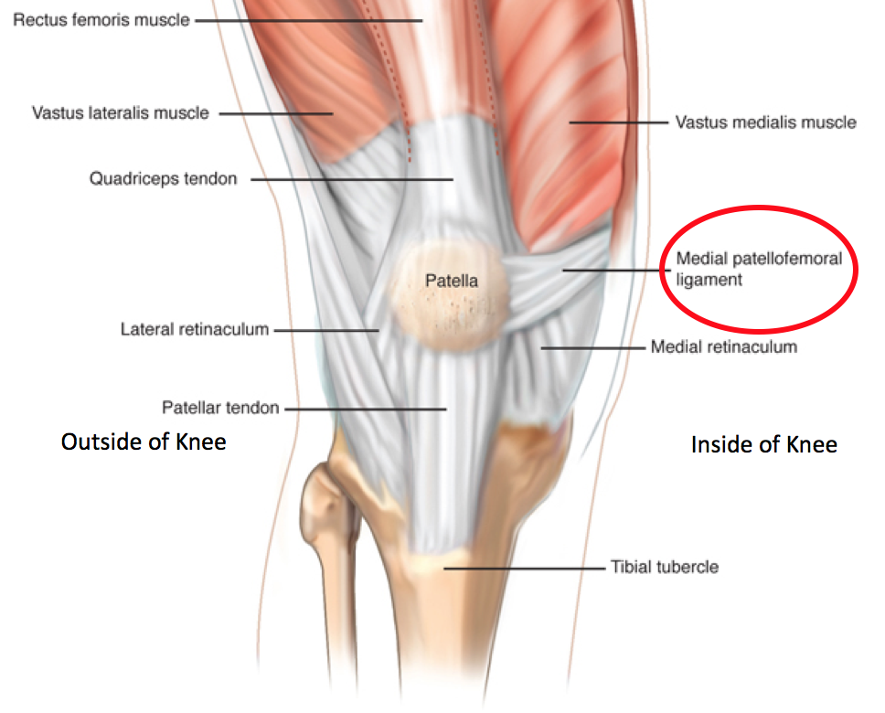
Above: The MPFL is circled in Red
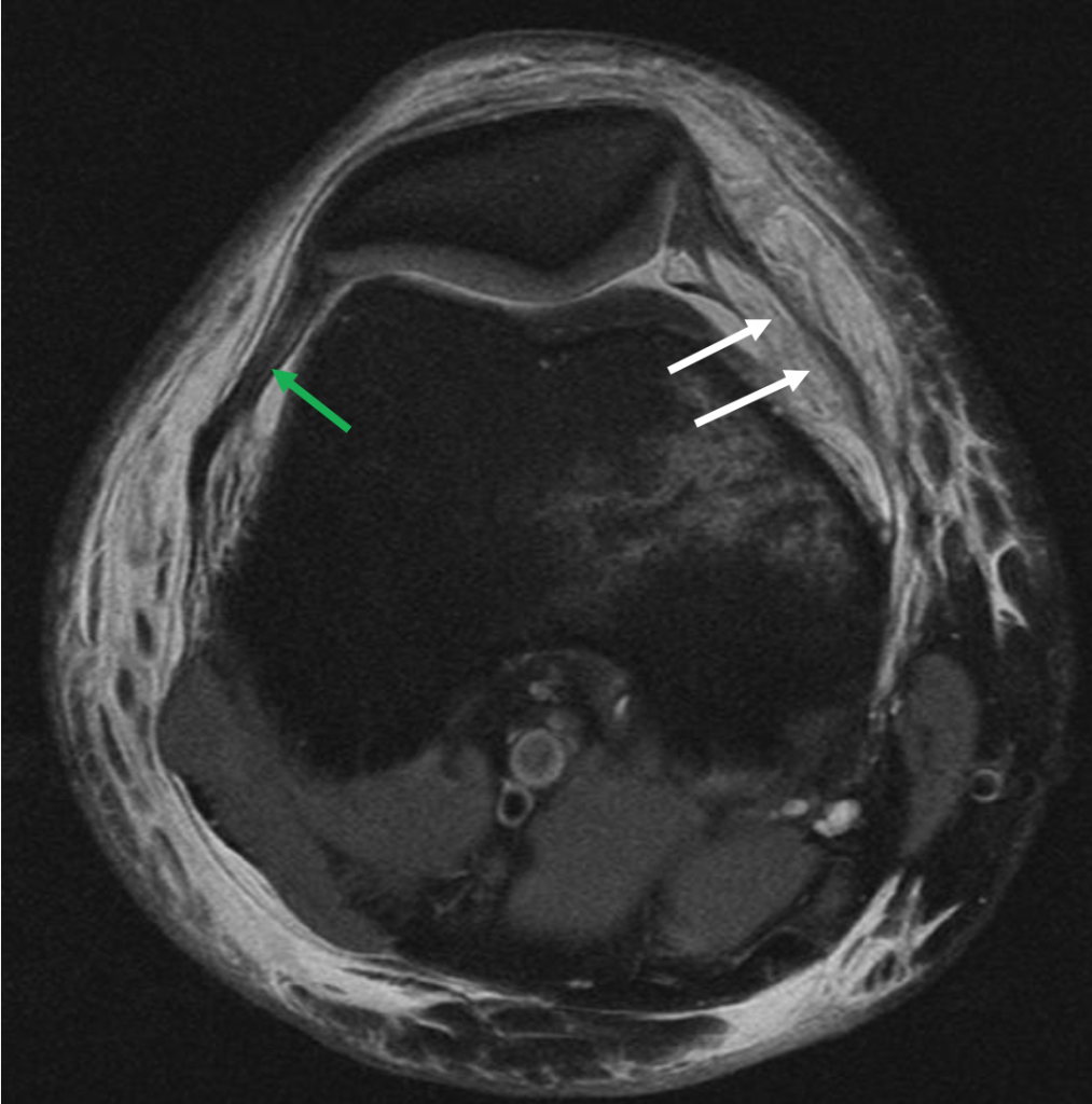
Above: White arrows indicate Ruptured MPFL, green arrow indicate a normal thick ligament (not ruptured)
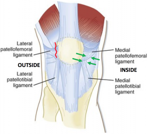
Lateral Release and Medial Tightening (Plication)
A Lateral Release is an arthroscopic (keyhole) procedure that is done to release the tight tissue on the outside of the knee that is pulling the kneecap out of joint. Medial tightening (plication is an operatio done through a small incision on the inside of the knee to tighten up the ligaments which pull the kneecap into joint (Medial Patellofemoral and Medial Patellotibial Ligaments).
Above: Green Arrow indicates Medial Plication, Red arrow indicates Lateral Release
Bone Procedures
Sometimes correction of the underlying bone is required to fix the mechanics which are driving the kneecap to dislocate. The major conditions which are treated with knee stabilisation surgery, are rotational problems in the femur (thighbone), height of the patella tendon or if the tendon which joins the kneecap to the tibia (shinbone) is positioned towards the outside, rather than being in the middle. If there is a significant underlying deformity and the bone is not considered, over time a purely soft tissue procedure may fail as the cause of the dislocation may not be addressed.
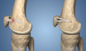
Tibial Tuberosity (Tubercle) Transfer
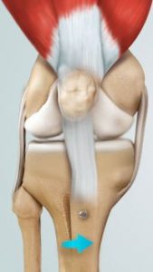
The tibial tuberosity is the part of the shinbone that the patella tendon inserts into. If it is positioned too far laterally (towards the outside) it can pull the kneecap out of joint. This type of patella stabilisation surgery involves an incision on the inside of the knee, the tibial bone is cut, and repositioned, then it is held in place with 2 or 3 screws.
De-Rotational Osteotomies
If abnormal rotation of the femur or tibia is contributing to the deforming forces upon the kneecap, then re-alignment of these bones may be required to alleviate these forces. This type of patellofemoral stabilisation is more major surgery, as it involves cutting the femur or tibia, and using plates or rods to hold them in place until healing occurs.
Above: Tibial Tuberosity Transfer with 1 Screw
Dr David Slattery
FRACS MBBS (Hons) LLB FAOrthA
Dr David Slattery is an orthopaedic surgeon based in Melbourne with over 10 years of experience, with a special focus on hip and knee joint preservation and replacement. With qualifications in both medicine and law, he brings a unique and comprehensive approach to patient care. His surgical techniques are minimally invasive and evidence-based, designed to reduce pain and enhance recovery.
Trained in leading institutions across Europe and the USA, Dr Slattery offers advanced treatments for a wide range of joint conditions. He is deeply committed to patient outcomes and takes pride in tailoring treatment plans to each individual. Whether you’re an athlete or seeking relief from chronic joint pain, his goal is to restore function and improve your quality of life.
To book an appointment please contact Dr Slattery’s rooms on
03 9819 6934

We aim to see all fractures within 24hrs