Click on the links below to find out more

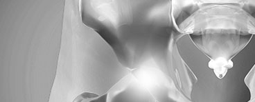
Perthes Disease (Legg-Calve-Perthes Disease)
Treatment
The goal of treatment is to relieve pain, restore normal hip movement and maintain a round ball and socket hip joint. If left untreated, the femoral head can deform which can lead to further hip problems in such as early onset arthritis.
There are many treatment options for Perthes disease. Factors when developing a treatment plan for your child, include:
- Your child’s age. Younger children (age 6 and below) have a greater potential for developing new, healthy bone.
- The degree of damage to the femoral head. If more than 50% of the femoral head has been affected by necrosis, the potential for regrowth without deformity is lower.
- The stage of disease at the time your child is diagnosed. How far along your child is in the disease process affects which treatment options your doctor will recommend.
Nonsurgical Treatment
Observation
For very young children (those 2 to 6 years old) who show few changes in the femoral head on their initial x-rays, the recommended treatment may be observation using x-rays to make sure the regrowth of the femoral head is on track as the disease runs its course.
Anti-inflammatory medications
Painful symptoms are caused by inflammation of the hip joint. Anti-inflammatory medicines, such as ibuprofen, are used to reduce inflammation, and these may be recommended by Mr Slattery for several months.
Limiting activity
Avoiding high impact activities, such as running and jumping, will help relieve pain and protect the femoral head. Crutches or a walker may be used to prevent your child from putting too much weight on the joint.
Physiotherapy exercises
Hip stiffness is common in children with Perthes disease and physiotherapy exercises are recommended to help restore and maintain hip joint range of motion. Parents or other caregivers are needed to help the child complete the exercises.
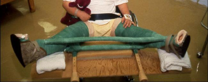
Petrie casts are two long-leg casts with a bar that hold the legs spread apart in a position similar to the letter “A.”
Casting and bracing
If range of motion becomes limited or if x-rays or other image scans indicate that a deformity is developing, a cast or brace may be used to keep the head of the femur in its normal position within the acetabulum.
Petrie casts are two long-leg casts with a bar that hold the legs spread apart in a position similar to the letter “A.”
Surgical Treatment
Arthrogram
Mr Slattery will inject a small amount of special dye into the hip joint then take a series of special x-ray images called arthrograms to see the degree of deformity of the femoral head and to make sure of the position of the femoral head the head accurately. In an arthrogram, a small amount of dye is injected into the hip joint to make the anatomy even easier to see.
Tenotomy
In some cases, the adductor longus muscle in the groin is very tight and prevents the hip from turning into the proper position. In this case, Mr Slattery may perform a minor procedure to release this tightness, called a tenotomy — before applying the casts.
Surgical Realignment
Surgical realignment of the bones (either femur(thighbone) or pelvis (hip bone)) is most often recommended when:
- Your child is older than age 8 at the time of diagnosis. Because the potential for deformity in older children, preventing damage to femoral head is even more critical.
- More than 50% of the femoral head is damaged. Keeping the femoral head within the rounded acetabulum may help the bone grow into a functional shape.
- Nonsurgical treatment has not kept the hip in correct position for healing.
The most common surgical procedure for treating Perthes disease is an osteotomy. In this type of procedure, the bone is cut and repositioned to keep the femoral head within the acetabulum. This alignment is kept in place with screws and plates, which will be removed after the healed stage of the disease.
In many cases, the femur bone is cut to realign the joint. Sometimes, the socket must also be made deeper because the head of the femur has actually enlarged during the healing process and no longer fits snugly within it. After either procedure, the child is usually placed in a cast for 6 to 8 weeks to protect the alignment.

An osteotomy of the femur places the femoral head in a better position to heal.
Outcomes
In most cases, the long-term prognosis for children with Perthes is good. However, if there is deformity in the shape of the femoral head, there is the potential for future problems. If the deformed head still fits into the acetabulum, problems may be avoided. In cases where the deformed head does not fit well into the acetabulum, hip pain or early onset of arthritis is likely in adulthood.

Dr David Slattery
FRACS MBBS (Hons) LLB FAOrthA
Dr David Slattery is an orthopaedic surgeon based in Melbourne with over 10 years of experience, with a special focus on hip and knee joint preservation and replacement. With qualifications in both medicine and law, he brings a unique and comprehensive approach to patient care. His surgical techniques are minimally invasive and evidence-based, designed to reduce pain and enhance recovery.
Trained in leading institutions across Europe and the USA, Dr Slattery offers advanced treatments for a wide range of joint conditions. He is deeply committed to patient outcomes and takes pride in tailoring treatment plans to each individual. Whether you’re an athlete or seeking relief from chronic joint pain, his goal is to restore function and improve your quality of life.
To book an appointment please contact Dr Slattery’s rooms on
03 9819 6934

We aim to see all fractures within 24hrs