Click on the links below to find out more


OSTEOCHONDRAL DEFECTs (OCD)
OVERVIEW
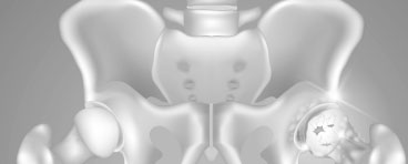
Osteochondral defects of the knee are a common source of knee pain, swelling and discomfort in children, adolescents and young adults. Osteochondral defects (OCDs) are diseased areas of bone (“osteo”) and cartilage (“chondral”). There are two main types of Osteochondral defects. Traumatic, which are normally as a result of patella dislocation or injury to the knee joint and Osteochondritis Dissecans, which occurs typically without a history of trauma and may affect both left and right knees.
ANATOMY
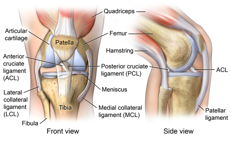
Normal knee anatomy is composed of the femur (thighbone), tibia(shinbone) and patella (kneecap). These are covered with a layer of cartilage which provides a smooth and slippery surface to allow movement through the knee joint. The cartilage is strongly adherent to the underlying bone, and it cushions the impact during walking and running. If healthy cartilage is damaged, a rough surface forms which causes the knee joint to degenerate and wear out (arthritis).
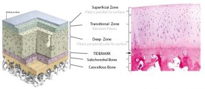
Normally the cartilage of the knee is strongly adherent to the underlying bone in layered ‘zones’ (above).

Dr David Slattery
FRACS MBBS (Hons) LLB FAOrthA
Dr David Slattery is an orthopaedic surgeon based in Melbourne with over 10 years of experience, with a special focus on hip and knee joint preservation and replacement. With qualifications in both medicine and law, he brings a unique and comprehensive approach to patient care. His surgical techniques are minimally invasive and evidence-based, designed to reduce pain and enhance recovery.
Trained in leading institutions across Europe and the USA, Dr Slattery offers advanced treatments for a wide range of joint conditions. He is deeply committed to patient outcomes and takes pride in tailoring treatment plans to each individual. Whether you’re an athlete or seeking relief from chronic joint pain, his goal is to restore function and improve your quality of life.
To book an appointment please contact Dr Slattery’s rooms on
03 5752 5020

We aim to see all fractures within 24hrs