Click on the links below to find out more
OSTEOCHONDRAL DEFECTs (OCD)
DESCRIPTION
Osteochondral defects of the knee are segments of the knee cartilage and bone which detaches from the underlying bone. This can cause pain, swelling, clicking, catching and locking of the knee. They typically affect young people, and more commonly men than women.
Osteochondral Defects
An osteochondral defect is comprised of varying amounts of bone (osteo) and cartilage (chondral). The depth of the OCD determines the quantity of bone and cartilage that are affected. Cartilage typically has very little healing potential, whereas bone heals well when it is in the correct position (eg undisplaced broken bones). Traumatic OCDs are typically associated with direct injuries or kneecap dislocations. These are usually displaced and do not heal by themselves.
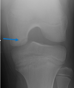
Osteochondritis Dissecans (OCDs without trauma) occur during childhood for unknown reasons. There is a spectrum of these lesions ranging from undisplaced lesions with intact cartilage to completely displaced and free floating loose pieces of bone and cartilage within the knee

Blue arrow indicates Osteochondritis Dissecans
Osteochondral defects can occur in a variety of locations within the knee, and they can also occur in other joints in the body, such as ankles, hips and elbows. The size, position and classification of the lesion will determine what treatment is indicated. This can range from observation, through to keyhole or open surgery. Please contact Dr Slattery to discuss what treatment is appropriate for your knee condition.

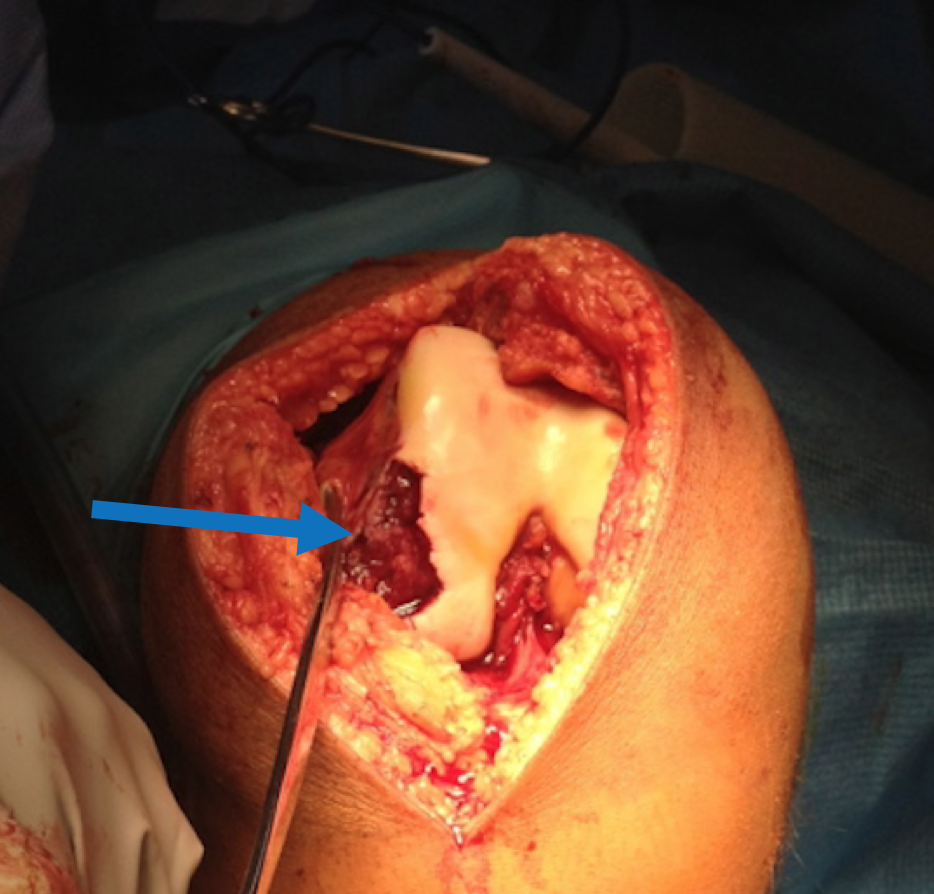
The blue arrow in the image above demonstrates the defect in the normal knee cartilage (shiny white) on the outside of the knee. This osteochondral fracture was as a result of a kneecap dislocation.

Dr David Slattery
FRACS MBBS (Hons) LLB FAOrthA
Dr David Slattery is an orthopaedic surgeon based in Melbourne with over 10 years of experience, with a special focus on hip and knee joint preservation and replacement. With qualifications in both medicine and law, he brings a unique and comprehensive approach to patient care. His surgical techniques are minimally invasive and evidence-based, designed to reduce pain and enhance recovery.
Trained in leading institutions across Europe and the USA, Dr Slattery offers advanced treatments for a wide range of joint conditions. He is deeply committed to patient outcomes and takes pride in tailoring treatment plans to each individual. Whether you’re an athlete or seeking relief from chronic joint pain, his goal is to restore function and improve your quality of life.


