Click on the links below to find out more


Hip- Avascular Necrosis/Osteonecrosis
DESCRIPTION
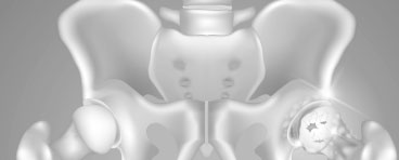
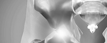
Avascular necrosis of the hip develops when the blood supply to the femoral head is disrupted. Without adequate nourishment, the bone in the head of the femur dies and gradually collapses. As a result, the articular cartilage covering the hip bones also collapses, leading to disabling arthritis.
Risk Factors
Although it is not always known what causes the lack of blood supply, there are a number of risk factors that can make it more likely for someone to develop the disease:
- Injury : Hip dislocations, hip fractures, and other injuries can damage the blood vessels and impair circulation to the femoral head
- Alcoholism
- Corticosteroid medicines : Many diseases, such as asthma, rheumatoid arthritis and systemic lupus erythematosus, are treated with steroid medications. Although it is not known exactly why these medications can lead to osteonecrosis, research shows that there is a connection between the disease and long-term steroid use.
- Other medical conditions : Osteonecrosis is associated with other diseases, including Caisson disease (diver’s disease or “the bends”), sickle cell disease, myeloproliferative disorders, Gaucher’s disease, systemic lupus erythematosus, Crohn’s disease, arterial embolism, thombosis and vasculitis
Symptoms
A osteonecrosis develops in stages. Hip pain is typically the first symptom. This may lead to a dull ache or throbbing pain in the groin or buttock area. As the disease progresses, it will become more difficult to stand and put weight on the affected hip, and moving the hip joint will be painful.
How long it takes for the disease to progress through these stages varies from several months to over a year. It is important to diagnose this disease early, because some studies show that early treatment is associated with better outcomes.
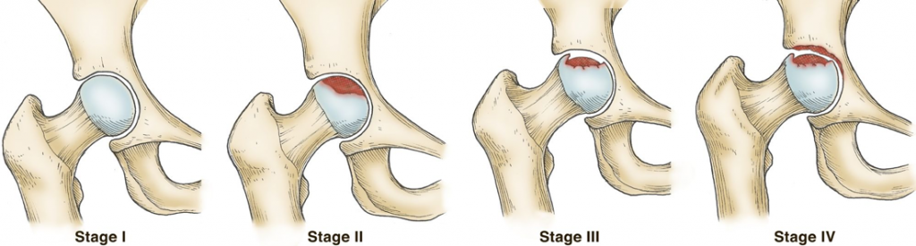
Osteonecrosis can progress from a normal, healthy hip (Stage I) to the collapse of the femoral head (Stage IV).
Imaging Tests
Imaging tests may help confirm a diagnosis and include:
X-Rays
These create pictures of dense structures, like bone. X-rays are used to determine whether the bone in the femoral head has collapsed and to what degree.
MRI Scans
Early changes in the bone that may not show up in an X-ray can be detected with an MRI scan. These scans are used to evaluate how much of the bone has been affected by the disease. An MRI may show early osteonecrosis that has yet to cause symptoms (for example, whether osteonecrosis is developing in the opposite hip joint).
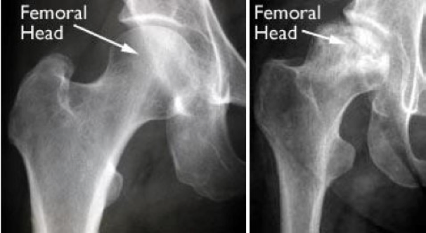
(Left) An X-ray of a healthy hip joint. (Right) An X-ray, where osteonecrosis has progressed to collapse of the femoral head.

Dr David Slattery
FRACS MBBS (Hons) LLB FAOrthA
Dr David Slattery is an orthopaedic surgeon based in Melbourne with over 10 years of experience, with a special focus on hip and knee joint preservation and replacement. With qualifications in both medicine and law, he brings a unique and comprehensive approach to patient care. His surgical techniques are minimally invasive and evidence-based, designed to reduce pain and enhance recovery.
Trained in leading institutions across Europe and the USA, Dr Slattery offers advanced treatments for a wide range of joint conditions. He is deeply committed to patient outcomes and takes pride in tailoring treatment plans to each individual. Whether you’re an athlete or seeking relief from chronic joint pain, his goal is to restore function and improve your quality of life.
To book an appointment please contact Dr Slattery’s rooms on
03 9819 6934

We aim to see all fractures within 24hrs