Click on the links below to find out more


Anterior Cruciate Ligament Injury (ACL)
DESCRIPTION
About half of all injuries to the anterior cruciate ligament occur along with damage to other structures in the knee, such as articular cartilage, meniscus, or other ligaments.
INJURY
The anterior cruciate ligament can be injured in several ways:
- Changing direction rapidly
- Stopping suddenly
- Slowing down while running
- Landing from a jump incorrectly
- Direct contact or collision, such as a football tackle
Several studies have shown that female athletes have a higher incidence of ACL injury than male athletes in certain sports. It has been proposed that this is due to differences in physical conditioning, muscular strength, and neuromuscular control. Other suggested causes include differences in pelvis and lower extremity (leg) alignment, increased ligament looseness, and the effects of estrogen on ligament properties.
SYMPTOMS
When you injure your anterior cruciate ligament, you might hear a “popping” noise and you may feel your knee give out from under you. Other typical symptoms include:
- Pain with swelling. Within 24 hours, your knee will swell. If ignored, the swelling and pain may resolve on its own. However, if you attempt to return to sports, your knee will probably be unstable and you risk causing further damage to the cushioning cartilage (meniscus) of your knee.
- Knee instability
- Loss of full range of motion
- Tenderness along the joint line
- Discomfort while walking
CLINICAL EVALUATION
Dr Slattery will take a detailed history of your injury, the symptoms you have had since, as well as your activity level and occupational demands. He will then perform a thorough physical examination of your knee to evaluate its stability, and the presence of other associated injuries. Specific tests for ACL rupture are:
- Lachmann Test
- Anterior Drawer Test
- Pivot Shift Test
It is also critical to evaluate the knee for its range of motion, swelling, alignment and for the presence of other ligament or meniscal injuries.
MEDICAL IMAGING (X Rays and MRI)
ACL injuries can be diagnosed both clinically (by your doctor) and with the help of imaging tests:
- X Rays: these can show fractures and the underlying alignment of your bones, which may need to be addressed. In children they also critically show the growth plates which need to be considered when doing surgical reconstruction.
- MRI Scans: these show an ACL rupture, and are very useful for assessing other injuries such as Meniscal Tears (up to 60%), Meniscal Avulsions, or Cartilage Damage.
- CT Scans: these can be used if there has been a prior ACL reconstruction to look at the position of the graft, and the tunnels
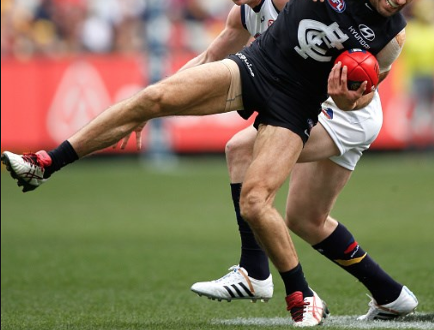
Above: Typical ACL Injury – Landing and twisting on a planted foot
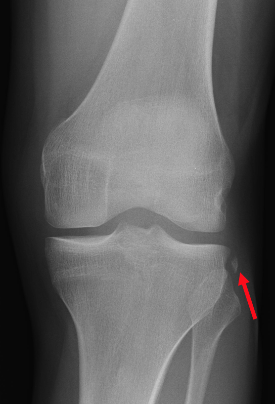
Above: X Ray showing a ‘Segond’ Fracture – indicating an ACL injury

Above: MRI Showing an ACL Rupture
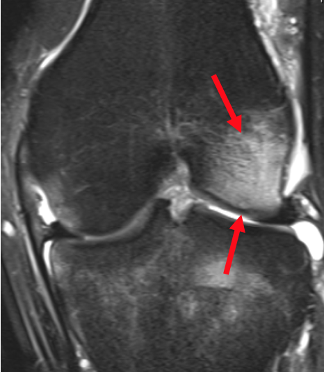
Above: MRI Showing bone bruising associated with an ACL injury

Dr David Slattery
FRACS MBBS (Hons) LLB FAOrthA
Dr David Slattery is an orthopaedic surgeon based in Melbourne with over 10 years of experience, with a special focus on hip and knee joint preservation and replacement. With qualifications in both medicine and law, he brings a unique and comprehensive approach to patient care. His surgical techniques are minimally invasive and evidence-based, designed to reduce pain and enhance recovery.
Trained in leading institutions across Europe and the USA, Dr Slattery offers advanced treatments for a wide range of joint conditions. He is deeply committed to patient outcomes and takes pride in tailoring treatment plans to each individual. Whether you’re an athlete or seeking relief from chronic joint pain, his goal is to restore function and improve your quality of life.
To book an appointment please contact Dr Slattery’s rooms on
03 9819 6934

We aim to see all fractures within 24hrs