Click on the links below to find out more


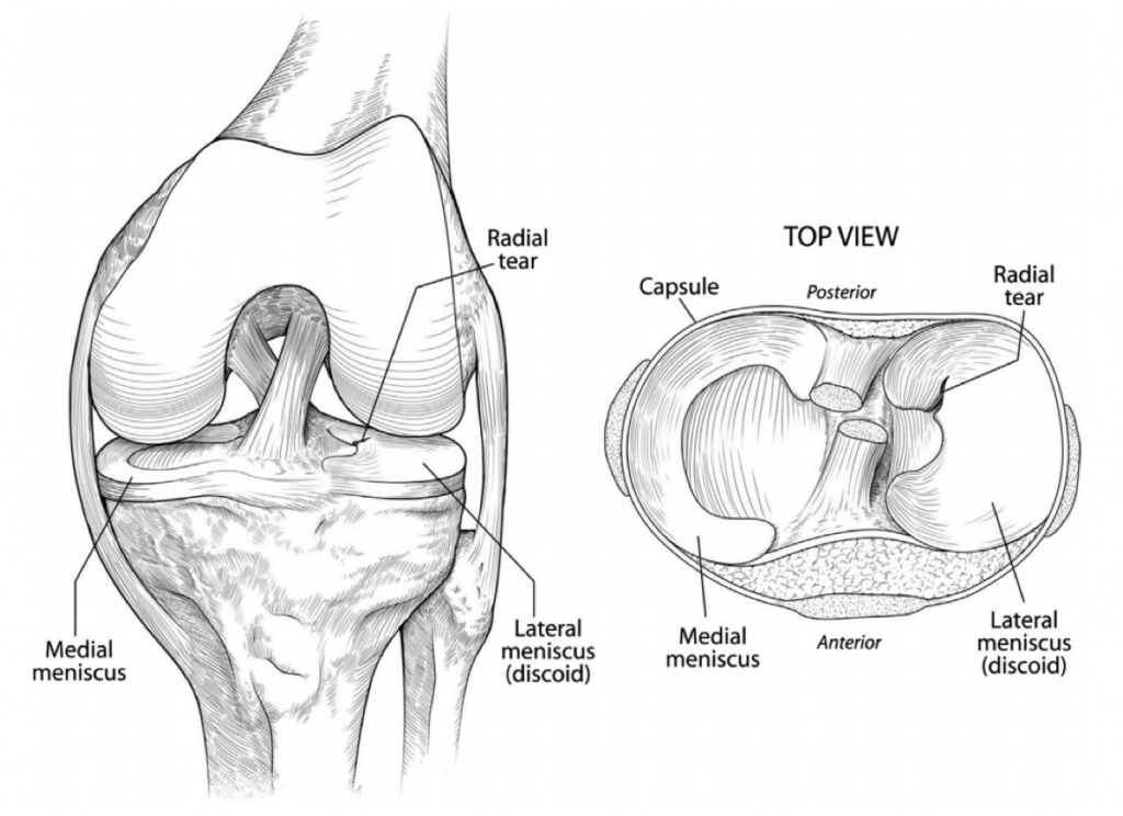
Discoid Meniscus
DESCRIPTION
A discoid meniscus is an abnormally formed meniscus (shock absorber) in the knee. It is more semi-circular rather than crescent shaped and has reduced stability compared to a normal meniscus.
Causes
The cause of a discoid meniscus is unknown. Proposed causes are due to a failure during embryological development, or that it is due to a lack of fixation to the tibia.
Symptoms
The symptoms of a discoid meniscus are highly variable. In some cases they may not have any symptoms at all, in the worst cases they can cause significant pain, swelling, discomfort and can cause the knee to ‘lock’ (get stuck and unable to move).
Commonly in young children, parents may notice a non-painful popping or clicking in the knee. In older children/adolescents they may feel as though the knee gives way, or if the meniscus has torn, they may have pain and swelling.
Investigations
Initial X rays can often be reported as normal, as the signs on X ray are quite subtle (see image at right).
On MRI, the charactertic finding is a “bow tie” sign present on multiple slices (see MRI on right)
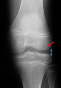
Above: X Ray showing increased lateral joint space (blue arrow) and a ‘squared off’ lateral condyle (red arrow)
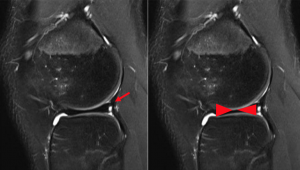
Above: MRI showing the ‘bow tie’ sign of a discoid lateral meniscus

Dr David Slattery
FRACS MBBS (Hons) LLB FAOrthA
Dr David Slattery is an orthopaedic surgeon based in Melbourne with over 10 years of experience, with a special focus on hip and knee joint preservation and replacement. With qualifications in both medicine and law, he brings a unique and comprehensive approach to patient care. His surgical techniques are minimally invasive and evidence-based, designed to reduce pain and enhance recovery.
Trained in leading institutions across Europe and the USA, Dr Slattery offers advanced treatments for a wide range of joint conditions. He is deeply committed to patient outcomes and takes pride in tailoring treatment plans to each individual. Whether you’re an athlete or seeking relief from chronic joint pain, his goal is to restore function and improve your quality of life.
To book an appointment please contact Dr Slattery’s rooms on
03 5752 5020

We aim to see all fractures within 24hrs