Click on the links below to find out more


Patellar Dislocation & Instability (Unstable Kneecap)
DESCRIPTION
There are a several ways in which the kneecap can become unstable or dislocate. In many cases, the patella dislocates with very little force because of an abnormality in the structure of the knee. A shallow or uneven groove in the femur can make dislocation more likely. Some people’s ligaments are looser, making their joints extremely flexible and more prone to patellar dislocation. This occurs more often in girls, and the problem may affect both knees.
Causes
Many things can cause patellar dislocation, such as a shallow groove in the femur or direct force on the knee joint.
In those with normal knee structure, patellar dislocations are often the result of a direct blow or a fall onto the knee. This incidence is more common in high-impact sports, such as football. Dislocations can occur without contact as well. An example is that of a right-handed baseball player who dislocates the right patella while swinging the bat. When the right foot is planted on the ground and the torso rotates during the swing, the patella lags behind, resulting in dislocation.

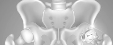
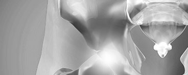
There are are a range of forces acting to cause the patella to dislocate (Left Image)
- Muscle imbalance (3 muscles act to pull it out, 1 to pull it in)
- ‘Q’ Angle: the angle of the muscle pull
- Femoral and Tibial Rotation – excessive rotation increases pressure pushing the patella out of its track
- Ligament Strength/Integrity: the MPFL (Medial Patellofemoral Ligament) is the primary ligament restraint, once torn it does not function properly
It is important that these are assessed to establish the cause of the dislocation, and to prevent its recurrence.
Symptoms
The symptoms associated with a patellar dislocation depend on how far out of place the patella has moved and how much damage occurred when it happened.
Some general symptoms include:
- Pain
- Feeling the kneecap shift or slide out of the groove
- Feeling the knee buckle or give way
- Hearing a popping sound when the patella dislocates
- Swelling
- A change in the knee’s appearance – the knee may appear misshapen or deformed
- Apprehension or fear when running or changing direction
Over time weakness of the knee will occur and patients will avoid activities which provoke dislocation to occur
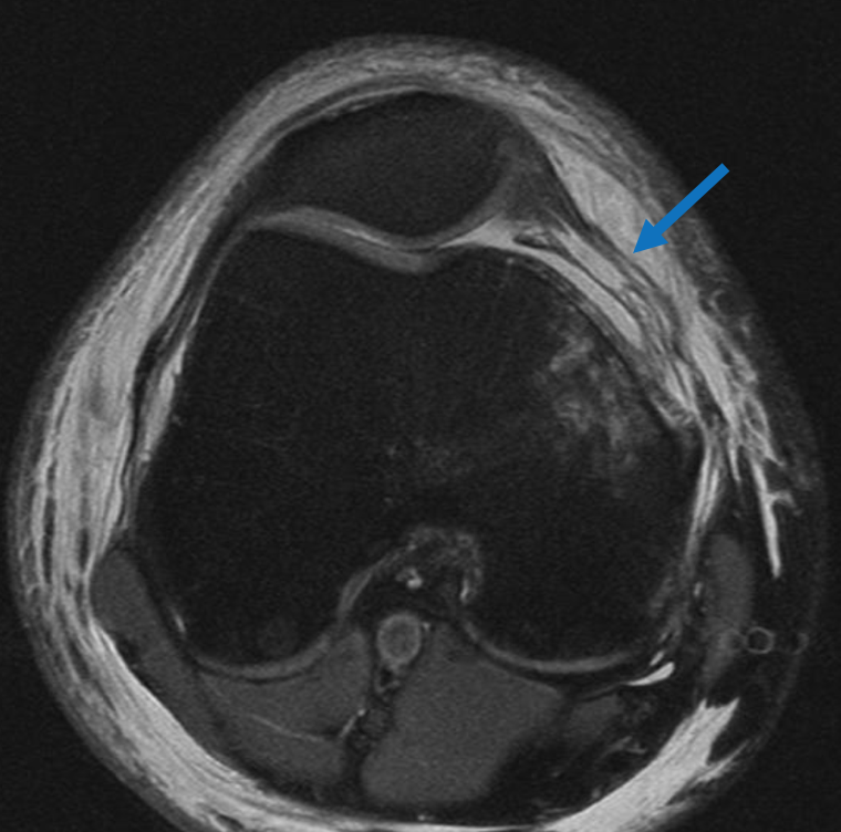
(Above) A MRI Scan demonstrating an MPFL rupture (Blue Arrow)
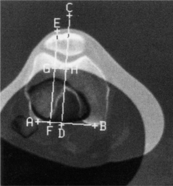
(Above) A CT Scan demonstrating the TTTG (Tibial Tuberosity to Trochlea Groove) distance – this is a key consideration when determining the cause of a patella dislocation.
Imaging Tests
X-rays
These tests create clear pictures of bone. These are useful to look for skeletal abnormalities in the knee which predispose to patella dislocation, such as a ‘high riding’ patella (‘patella alta’), or a shallow groove for the patella to run in (shallow trochlea). They can also identify loose bodies and fractures which can be the result of a dislocation.
Magnetic resonance imaging (MRI) scans
These scans create better pictures of the soft structures surrounding the knee, like ligaments. An MRI is very useful in assessing the cartilage and ligaments, which are commonly damaged during patella dislocation
CT Scans
These are very useful for looking at a patients rotation and underlying bone structure, which can be a significant contributing factor in patellar dislocation.

Dr David Slattery
FRACS MBBS (Hons) LLB FAOrthA
Dr David Slattery is an orthopaedic surgeon based in Melbourne with over 10 years of experience, with a special focus on hip and knee joint preservation and replacement. With qualifications in both medicine and law, he brings a unique and comprehensive approach to patient care. His surgical techniques are minimally invasive and evidence-based, designed to reduce pain and enhance recovery.
Trained in leading institutions across Europe and the USA, Dr Slattery offers advanced treatments for a wide range of joint conditions. He is deeply committed to patient outcomes and takes pride in tailoring treatment plans to each individual. Whether you’re an athlete or seeking relief from chronic joint pain, his goal is to restore function and improve your quality of life.
To book an appointment please contact Dr Slattery’s rooms on
03 9819 6934

We aim to see all fractures within 24hrs