Click on the links below to find out more


Acetabular Retroversion
DESCRIPTION
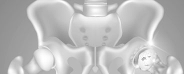
Acetabular retroversion is a condition where the hip socket (acetabulum) faces backwards (retroversion) rather than forwards.
Cause
Acetabular retroversion is a form of hip dysplasia (where the hip fails to form normally). There is no known cause for acetabular retroversion, however it may commonly exist with other hip problems such as FAI (femoroacetabular impingment), SCFE, and Perthes Disease.
Clinical features of Acetabular Retroversion
In many cases Acetabular Retroversion may be an incidental finding noticed on X-rays or scans without any problems at all. However, some patients may develop symptoms due to the femur (thighbone) abnormally hitting the pelvis, or instability from the femoral head slipping backwards out of the hip joint.
Common symptoms include:
- Hip Pain
- Hip Clicking/Catching
- Hip Giving way
- Limping
- Fatigue
- Stiffness
Investigations
In many cases Acetabular Retroversion may be missed on plain X-rays. It is recommended that Specialist Musculoskeletal Radiologists, or Specialist Hip Surgeons assess x-rays looking for Acetabular Retroversion. The signs on X-rays are often subtle and easily missed, and good quality X-rays are essential for the diagnosis.
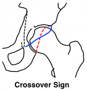
Signs on X-ray include:
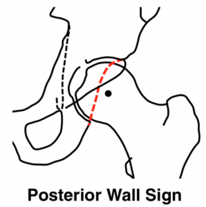
- Posterior Wall Sign
- Ischial Sign Sign
- Crossover Sign
CT Scans and MRI scans are very useful as they give a cross – sectional picture of the acetabulum and are the most accurate for looking at the bone and associated soft tissue structure of the hip joint.
Specific MRI and CT scans are useful for assessing the health of the hip cartilage, as well as the rotation of the entire leg which can contribute to the symptoms that patients with acetabular retroversion may experience.
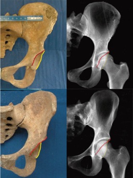
Top: The normal appearance of the Acetabulum (Cup), showing the front (Red/anterior) and back (Yellow/posterior) walls. Bottom: Acetabular Retroversion where the front wall is more exposed than the back wall. Jamali et al 2007.
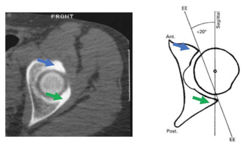
Above: CT Scans are very useful for looking at the acetabular position – in cross section above the blue arrow is the front wall and the green arrow is the back wall.

Dr David Slattery
FRACS MBBS (Hons) LLB FAOrthA
Dr David Slattery is an orthopaedic surgeon based in Melbourne with over 10 years of experience, with a special focus on hip and knee joint preservation and replacement. With qualifications in both medicine and law, he brings a unique and comprehensive approach to patient care. His surgical techniques are minimally invasive and evidence-based, designed to reduce pain and enhance recovery.
Trained in leading institutions across Europe and the USA, Dr Slattery offers advanced treatments for a wide range of joint conditions. He is deeply committed to patient outcomes and takes pride in tailoring treatment plans to each individual. Whether you’re an athlete or seeking relief from chronic joint pain, his goal is to restore function and improve your quality of life.
To book an appointment please contact Dr Slattery’s rooms on
03 9819 6934

We aim to see all fractures within 24hrs